Figure 1.
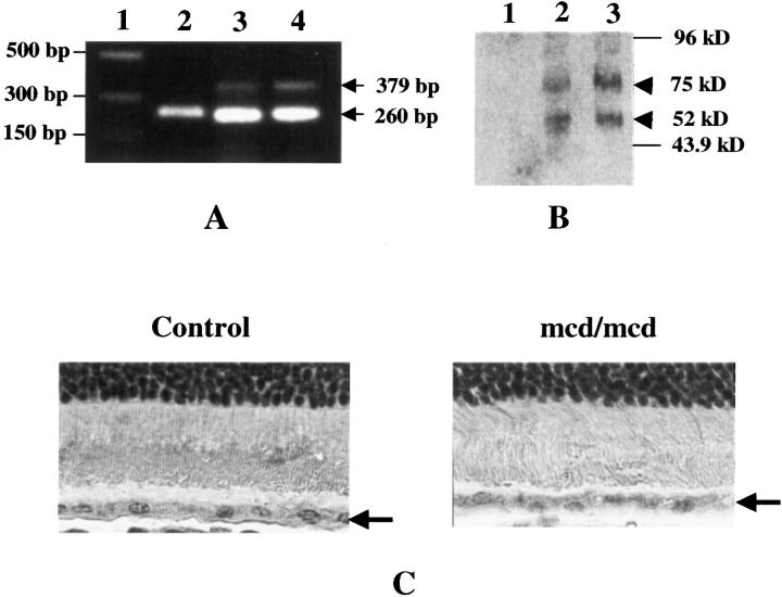
Molecular characterization of the mcd/mcd mouse. A: RT-PCR of 12-month-old mcd/mcd, mcd, and C57BL/6 mice retinas showing presence of the 379-bp CatDm1 cDNA (arrow) and 260-bp HPRT cDNA (arrow) fragments. Lane 1, DNA marker; lane 2, C57BL/6; lane 3, mcd; lane 4, mcd/mcd. B: Western blot analysis of mouse retinae using a monoclonal anti-human CatD mAb. Lane 1, C57BL/6; lane 2, mcd; lane 3, mcd/mcd eyes. Human proCatD appeared at 52 kd in mcd and mcd/mcd eyes (arrowhead). Note: the appearance of a higher MW complex at 75 kd (arrowhead). C: Immunohistochemistry of human CatD using a monoclonal anti-human CatD mAb. CatD-positive signal was detected in the RPE cell layer (arrow) of the mcd/mcd mouse eye but not in the age-matched C57BL/6 eye. Original magnifications, ×1000.

