
Figure 4.
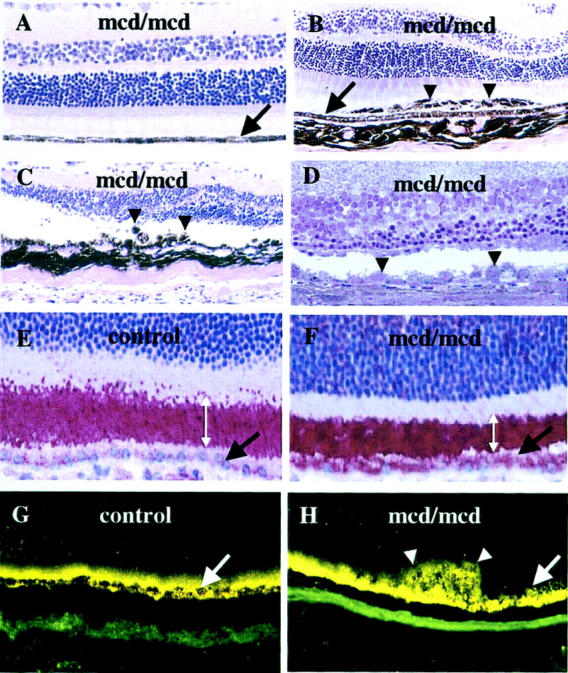
Light microscopy of 12-month-old mcd/mcd (A–C and E–H) and C57BL/6 (A–C and E–H) mouse and an 18-month-old mcd/mcd (D) retinae. The RPE layer in each figure is indicated by a black or white arrow. A: Morphology of the normal region of an mcd/mcd mouse retina. B: Morphology of mcd/mcd mouse retina showing abnormal layers of pigmented cells (arrowheads). C: Abnormal hyper- and hypopigmented RPE cells (arrowheads) in mcd/mcd mouse retina and thinning of photoreceptor layer above the abnormal RPE cells. D: Toluidine blue-stained resin section showing abnormal RPE cells (arrowheads) and thinned photoreceptor cell layers (note: the area centralis is on the left side of the micrograph) (original magnification, ×200). E: ROS-immunohistochemistry of C57BL/6 mouse retina. F: ROS-immunohistochemistry of mcd/mcd mouse retina (note the shortening of the ROS and ROS-immunoreactivity localized in the RPE cells). Width of ROS is indicated by double-headed white arrows. G: Fluorescent microscopy of retinas of C57BL/6. H: mcd/mcd mouse. Note the higher fluorescent signal in the RPE cells and in the clumped cells (white arrow) (original magnification, ×400).