
Figure 5.
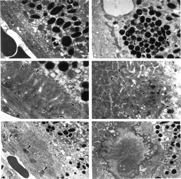
Electron micrographs of an 18-month-old mcd/mcd mouse retina. A: Electron micrograph of a relatively normal segment of the RPE cell layer in an as yet unaffected region of the mcd/mcd mouse. The basal infoldings are clearly seen (arrows). B: An RPE cell from the retina of the mcd/mcd mouse. Note the large focus of poorly melanized granules in the cytoplasm (arrows). C: An electron micrograph from an affected area of the mcd/mcd mouse. A subepithelial deposit consisting of filaments, granules, and osmiophilic matrix can be seen between the RPE plasma membrane and its basal lamina. Note the dissection of the deposit by attenuated processes from the RPE cell (arrows). Bundles of filaments can be observed within the deposit (arrowheads). D: In a similar area to C the deposit contains bundles of filaments (small arrows), vesicles (arrowheads), granules, and widely spaced collagen (large arrow). E: Another subepithelial deposit containing membranous whorls (arrows). BM appears to be slightly thickened and vacuoles can be seen beneath the basal lamina of the RPE cells (arrowheads). F: An electron micrograph of a large spherule in the mcd/mcd mouse retina. Electron dense spicules can be seen around the periphery of this inclusion (arrows). Original magnifications: ×11,600 (A); ×9900 (B); ×17,300 (C); ×12,720 (D); ×6100 (E); ×8800 (F).