
Figure 1.
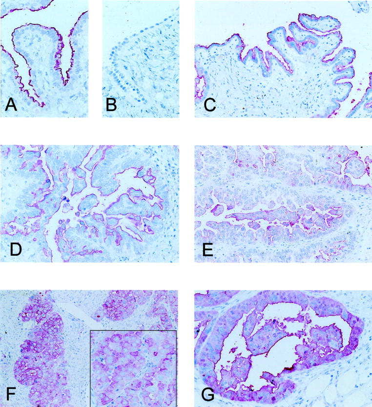
CD24 Immunohistochemistry. A: Physiological salpingeal mucosa with a strong luminally polarized membranous staining. B: Ovarian surface epithelium without any signal. C and D: Membranous staining without cytoplasmic reactivity in a borderline tumor of the ovary (C) and a serous carcinoma (D). E: Serous ovarian carcinoma with a weak cytoplasmic staining. F and G: Two invasive ovarian carcinomas with strong cytoplasmic staining, which is in part circumferentially accentuated.