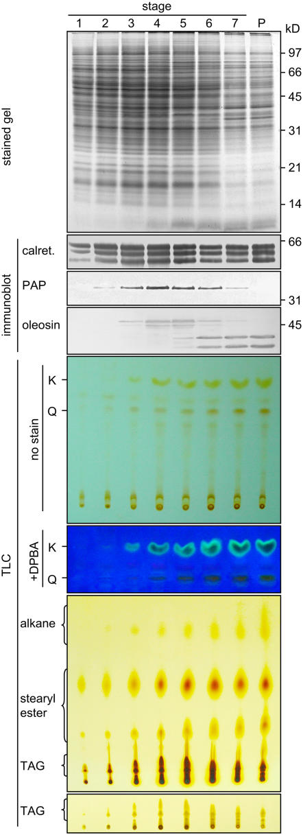
Figure 1.
SDS-PAGE, Immunoblot, and TLC Analyses of Extracts of Brassica Anthers of Seven Developmental Stages and Mature Pollen.
The various extracts applied to the gels or TLC plates represented those from an equal number of anthers. Gels were stained for proteins or subjected to immunoblot analyses with antibodies against calreticulin, elaioplast PAP, or tapetosome oleosin. The two major oleosins of 48 and 45 kD were converted to fragments of 37 and 35 kD, respectively, during anther development; the fragments were more immunoreactive than the originals (Ting et al., 1998). Positions of markers for polypeptide molecular masses are shown at right. The TLC plate after separation of deglycosylated flavonoids was photographed directly (full plate shown) or after being treated with DPBA and placed on top of a UV irradiation source (portion of the plate shown). K and Q denote kaempferol and quercetin, respectively. The TLC plate for lipid analyses was stained with iodine for 24 h to reveal all lipids, including the less reactive alkanes (full plate shown), or for 0.5 h to reveal TAGs more clearly (portion of the plate shown).

