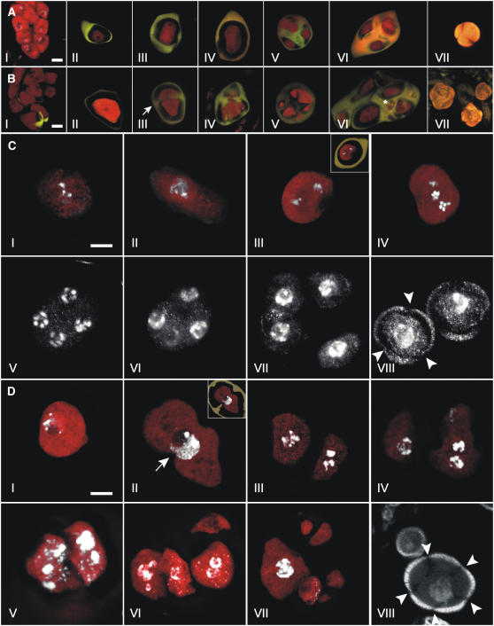
Figure 7.
Male Meiosis in Wild-Type and T161D Plants.
Confocal laser scanning microscopy of developmental stages of male meiosis in wild-type ([A] and [C]) and T161D ([B] and [D]) plants.
(A) and (B) Propidium iodide (PI; red) and aniline blue (AB; yellow) stains were used to paint meiotic cells and callose walls, respectively.
(A) I, wild-type pollen mother cells before callose deposition (triple stain with PI/AB/DAPI). II, premetaphase I. III, telophase I. IV, telophase II. V, early tetrad stage. VI, late tetrad stage. VII, released haploid uninucleate microspore. Bar = 5 μm.
(B) I, T161D pollen mother cells. Callose deposition has started in the bottom part of the micrograph. II, premetaphase I. III, dyad undergoing cytokinesis and deposition of callose between cells (arrow), presumably after telophase I. IV, continued callose wall deposition. V, heptad. Note different compartment sizes. VI, late-stage polyad with 11 nonuniform cellular compartments. Note division in the small compartment (asterisk). VII, released T161D microspores. Bar = 5 μm.
(C) and (D) PI (red) and DAPI (white) stains were used to visualize meiotic cells and DNA, respectively.
(C) Male meiosis in the wild type. I, pachytene. II, early diplotene. III, metaphase II. The inset depicts the same micrograph in overlay with callose deposition visualized by AB staining. IV, anaphase II. Two spindle poles reside in different focal planes in the center of the cell. V, early telophase II showing five chromatids at each spindle pole. VI, cytokinesis divides the nuclei at each pole of the second meiotic spindle. VII, tetrad stage. Callose is deposited between the four haploid meiotic products. VIII, released haploid uninucleate microspores with three pores (arrowheads). Bar = 5 μm.
(D) Male meiosis in T161D. I, meiocyte from T161D in pachytene/early diplotene. II, premetaphase I meiocyte. Note invaginations in the meiotic cell (arrow). The inset depicts the same micrograph in overlay with callose deposition visualized by AB staining. Note that callose projects into the meiotic cell invaginations. III to V, T161D dyads after cytokinesis and callose deposition. III, the left nuclei appear to be in anaphase/metaphase. IV, in the right cell, nuclear division appears to have taken place. V, cytokinesis appears to have taken place between the two nuclei in the left cellular compartment. VI, callose deposition has taken place between cells in a T161D tetrad. Note two prominent nuclei in the bottom left cellular compartment and no nuclei in the top left cell. VII, heptad with nonuniform cells embedded in callose. Note the large cell at left with large nuclei. The other cellular compartments appear to be enucleate. VIII, released T161D microspores with multiple pores (arrowheads). A large and a small microspore are shown. The exine layer appears normal in all sizes. Bar = 5 μm.

