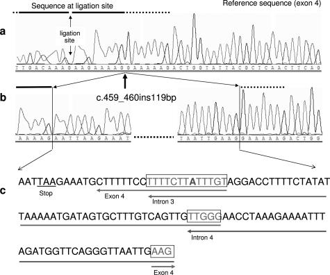
Figure 1.
Partial sequencing pattern of exon 4 and the inserted fragments. a: Normal sequence of APC exon 4. The sequence at the ligation site of MLPA probes for exon 4 (according to MRC Holland) is marked by a horizontal bar. b: Partial sequence of the mutant allele showing the breakpoint in exon 4 (arrow) and the sequence of the 5′ and 3′ end of the 119-bp insertion. c: Complete sequence of the 119-bp insertion in exon 4. The origin of the first 12 bp is unknown. The corresponding genomic origin of the other fragments is marked by horizontal arrows. The framed regions indicate the overlapping sequences at the ends of each fragment [one mismatched nucleotide (A) is shown in bold].

