
Fig. 1.
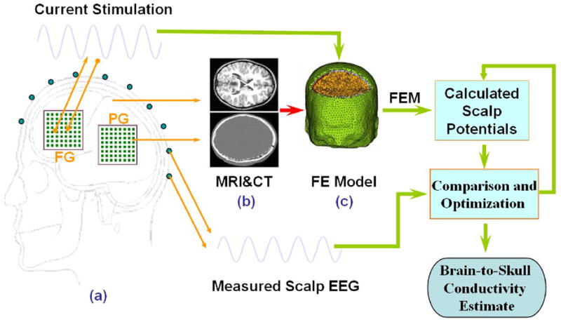
Schematic diagram of the procedures to estimate the in vivo brain-to-skull conductivity ratio in human subjects. The electrical potentials over the scalp are recorded during injection of current onto a pair of adjacent subdural electrodes (1 cm apart). The realistic geometry inhomogeneous head model was constructed from the MRI and CT images of the subject, including the brain, skull, scalp, and the silastic subdural grids. The finite element method is used to solve the electric fields due to current injection with various conductivity profiles. The difference between the measured and model-predicted scalp electric potentials is minimized by means of the simplex method, with the parameter of brain-to-skull conductivity ratio.