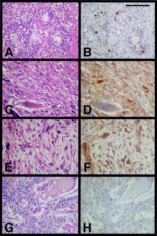
Figure 6.
H&E and immunohistochemical staining using the anti-SSX JOY-1 polyclonal antibody. A and B: Immature human testis from a 2-year-old boy with congenital heart disease. Seminiferous tubule exhibited intratubular staining of early spermatogenic cells, mainly spermatogonia. Neither Sertoli cells, interstitial cells, nor maturated spermatogenic cells were stained with JOY-1. C and D: Metastatic osteosarcoma of lung. E and F: MPNST. G and H: Synovial sarcoma containing a SYT-SSX2 fusion transcript. Heterogeneous SSX nuclear staining was found in osteosarcoma and MPNST, whereas no staining was observed in synovial sarcoma. Scale bar, 100 μm.

