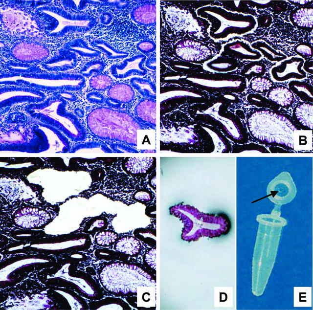
Figure 1.
Laser microdissection. A: A periodic acid-Schiff-stained section used to identify the degree of dysplasia. B, C, E: The laser cut line surrounds the dysplastic cells (B); these cells were harvested in the cap of a microtube (C, E). D: The tissue section after laser microdissection: DNA contamination by stromal and inflammatory cells surrounding the desired cells was avoided.

