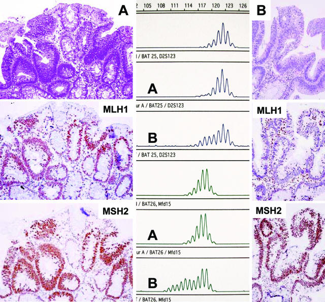
Figure 3.
Carcinoma arising in an adenoma in a case with partial loss of MSH2 in the adenoma. A: Focus of MSH2 expression in the adenoma (->, retained MSH2 immunostaining; AD, adenoma; CA, carcinoma; N, normal mucosa; * , lymphoid follicle as internal positive staining control). B: Enlarged inset from A with focal MSH2 staining and C shows the expression of MLH1 within the same area. D and E: MSI at BAT40 (D) and BAT25 (E) of MSH2-negative cells (D: lane 1, normal; lanes 2 and 3, adenomatous tissue without MSH2; lanes 4 and 5, with preserved MSH2 expression; E: lane 1, normal; lane 2, adenomatous tissue with MSH2 staining; and lanes 3 and 4, without MSH2 staining).

