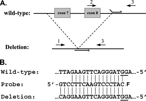
Figure 1.
Strategy. A: The positions of the primers (arrows) and probe on the wild-type and mutant CLN3 gene are shown. The fluorophore on the probe is indicated with an asterisk; not drawn to scale. B: The sequence of the probe is shown in between the complementary sequences of the wild-type and deletion alleles, with vertical lines connecting the base-paired residues. The –F on the 3′ end of the probe indicates the 6-FAM fluorophore and shows its position with respect to the G residues on the opposite strand. The probe is fully base-paired with the mutant sequence, but has three unmatched nucleotides at the 5′ end when annealed to a wild-type amplicon. The G residues that contribute to the quenching of the fluorescent signal are underlined in the normal and mutant sequences.

