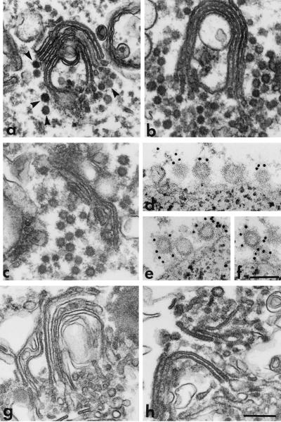
Figure 3.
Electron microscopic analysis of the effects of anti-P200 and anticoatomer antibodies on post-Golgi vesicle production. (a–c) Golgi membranes were incubated in reaction mixtures containing GMP-PNP and intact LCP, without (a), or with either anti-P200 (b) or anticoatomer antibodies (c), or with LCP that had been treated for immunodepletion with anti-P200 (g) or anticoatomer antibodies (h) linked to PGA beads. After incubation at 37°C (a–c), or at 20°C (g and h), the Golgi cisternae were recovered by centrifugation (10,000 × g for 10 min) and processed for electron microscopy. Note that in the control sample (a), despite extensive vesicle release (see Fig. 2C, ○—○), some coated vesicles (arrowheads) still remain associated with the Golgi cisternae. Although the biochemical experiments show that in b and c vesicles are not released or released to a very limited extent (see Figs. 2C and 6C, •—•), the electron micrographs show that coated vesicles are still produced but in the presence of the antibodies they form large aggregates that remain associated with the Golgi membranes. Immunodepletion with either anti-P200 (g) or anticoatomer (h) antibodies renders LCP incapable of supporting the formation of nonclathrin coated vesicles. (d–f) Vesicles released as in a were captured on magnetic beads containing immobilized anti-P200 antibodies. The beads were then incubated with the EAGE anticoatomer antibody followed by protein A gold (10 nm). Portions of the surface of beads are shown. Note that vesicles bound to the beads via the anti-P200/myosin II antibody are also labeled with the anticoatomer antibody (gold particles). Bars = 0.25 μm (a, b, c, g, and h); 0.1 μm (d–f).

