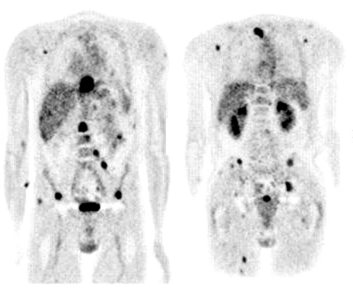
Figure 2.
A 57 year old man with oesophageal cancer and clinical stage T3N1 presented for staging evaluation. (Left) Anterior coronal and (right) posterior coronal FDG PET show multiple foci of intense metabolic activity compatible with widespread metastases. CT scan confirmed many of these sites, but was unable to detect spread to lymph nodes and distant soft tissue site.

