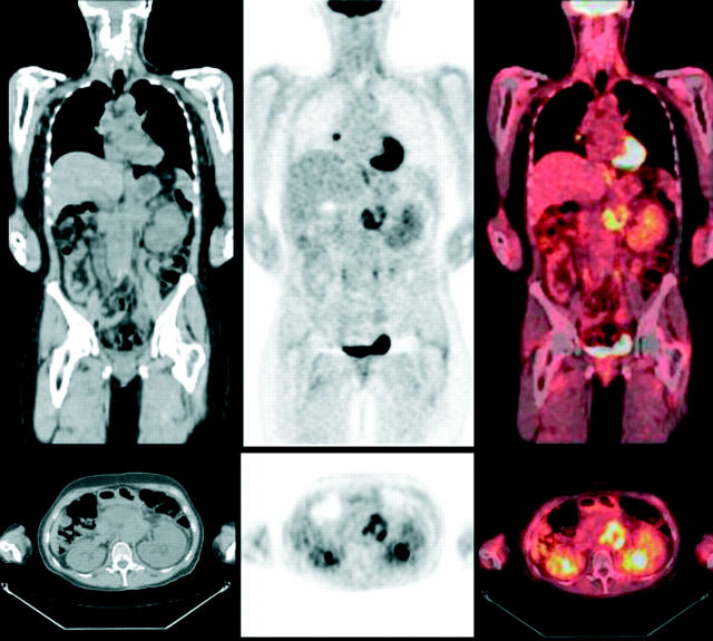
Figure 3.
A 43 year old woman with pancreatic cancer presented for evaluation of lung nodules. (Top row) Coronal and (bottom row) transaxial tomographic slices. (Left) CT shows the mass in the abdomen, but is unable to characterise the hilar lymph node. (Centre) FDG PET shows intense activity in the abdominal mass and increased uptake in the hilar lymph node (additional lung abnormalities not shown) compatible with local recurrence and distant metastases. Clinical follow up of lung abnormalities was consistent with metastases. (Right) Fusion of PET and CT localises the abnormalities.

