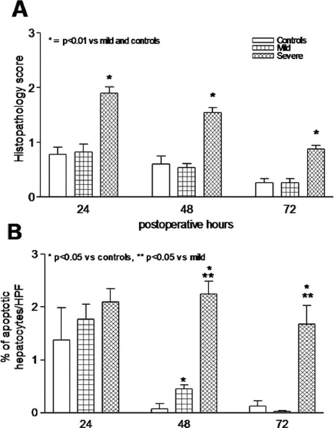
FIGURE 4. Histopathology scores in hematoxylin and eosin-stained liver tissue (A) after PH was significantly increased in severe steatotic rats compared with no changes between the control and mild steatotic rats (P < 0.01). The rate of apoptotic cells (B) remained increased in the severe steatotic rats compared with decreasing rates in the control and mild steatotic rats after PH. Values are mean ± SEM. *P < 0.05 versus controls. **P < 0.05 versus the mild steatosis group.
