
Figure 5.
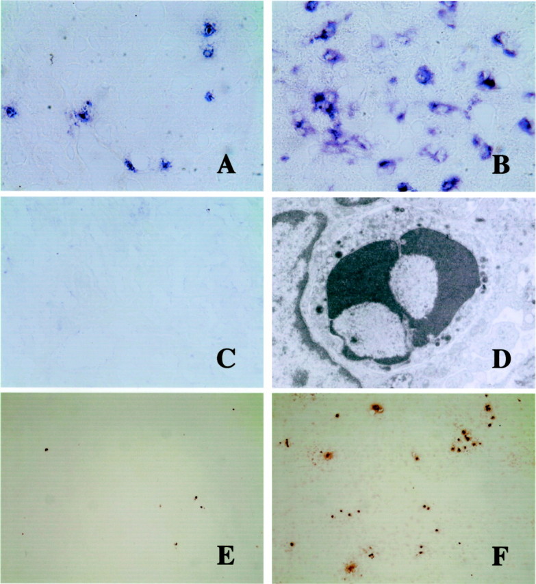
AIM mRNA expression in the liver by in situ hybridization (A-C), electron micrograph of an apoptotic lymphocyte (D), and TUNEL-staining of apoptotic cells (E and F). A–C: AIM mRNA is weakly expressed in a few round or spindle cells along the hepatic sinusoids of untreated AIM+/+ mice (A). At day 17 after C. parvum injection, AIM expression is augmented in macrophages (B), especially in granulomas. However, signal-positive cells were not detected in the liver of C. parvum-injected and non-injected AIM−/− mice (C). Original magnification, ×400. D: Electron micrograph of a lymphocyte in the granuloma with small electron-dense intracytoplasmic granules. Condensed nucleus suggests apoptosis of this granular lymphocyte. Original magnification, ×12,000. E and F: At 17 days TUNEL-labeled cells are more abundant in the liver of AIM−/− mice (F) than in AIM+/+ mice (E). Original magnification, ×40.