
Figure 2.
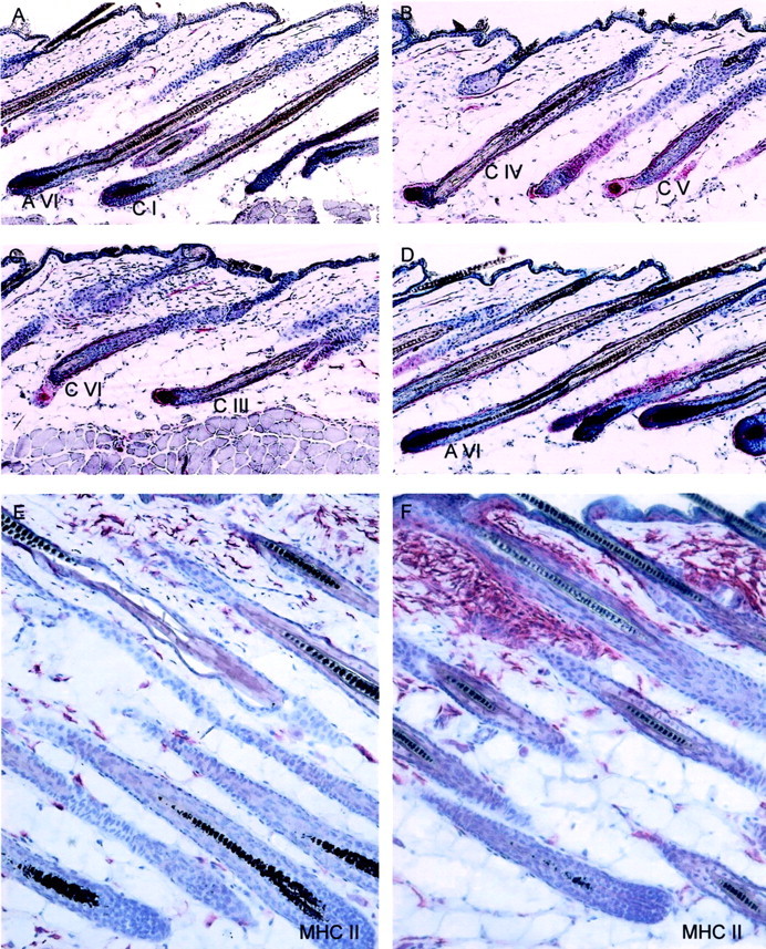
The effect of stress on the hair cycle stage is depicted in A–D. A: A representative area of control mice 16 days after depilation with the majority of HFs in anagen VI (AVI). B mirrors the effect of stress on the hair cycle stage on day 16 after depilation with HFs in catagen IV (CIV) or catagen V (CV). C: HF of stressed mice that received injection of SP, which mimicked the effect of stress on the vulnerability of HFs toward catagen progression with HFs in catagen III to VI (CIII-VI). D: A representative example of mice exposed to stress and treated with the NK1-RA, in which the majority of HFs were scored as anagen VI, similar to the nonstressed control group. E–F: The effect of stress on immunocytes in murine skin. E: Distribution of MHC-II+ cells in parafollicular dermis of nonstressed mice or in similar distribution in stressed mice that received the NK1-RA (bright red staining), compared to F, increased number of MHC-II+ cells in the bulge region, forming cluster after stress exposure. Similar staining patterns were present after injection of SP. Original magnifications, ×200.