
Figure 1.
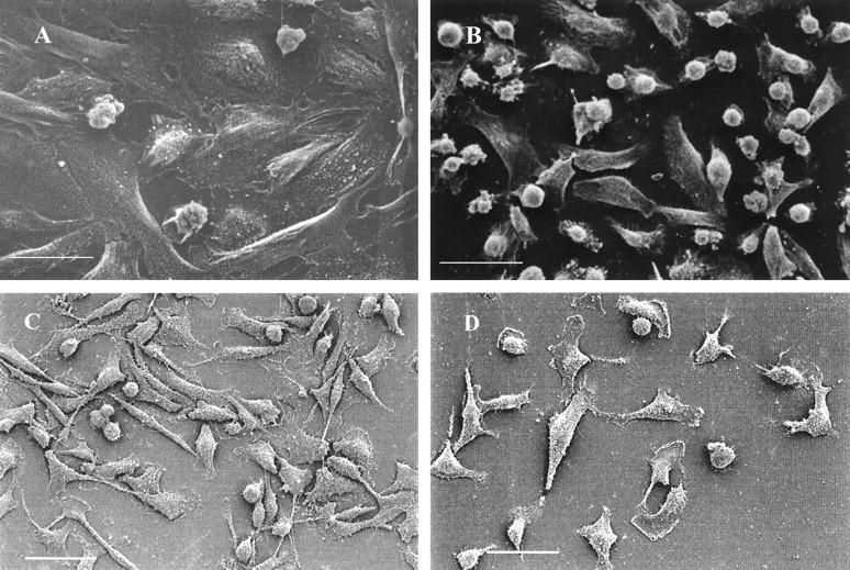
Scanning electron micrographs of progesterone-treated ABC28 (PR-transfected MDA-MB-231) cells (A) which are more flattened than vehicle- (0.1% ethanol) treated ABC28 cells (B) after 48 hours of treatment. Bar, 30 μm. Progesterone-treated and vehicle-treated MDA-MB-231 cells transfected with the pSG5 plasmid (C and D, respectively) looked similar to vehicle treated ABC28 cells. Bar, 50 μm.