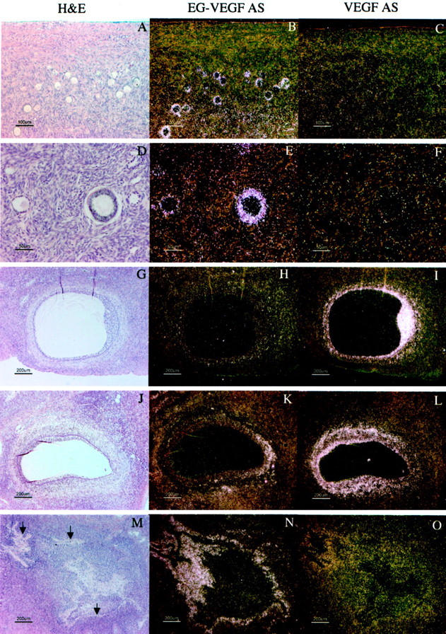
Figure 1.

VEGF and EG-VEGF expression in maturing follicles in normal ovaries. A–C: Primary and primordial follicles show strong expression of EG-VEGF (B) but little or no expression of VEGF (C). D–F: Maturing secondary follicles with multiple layers of granulosa cells maintain strong EG-VEGF expression, but show weak to moderate VEGF expression. G–I: Antral follicle (see arrowhead in Figure 5B ▶ ), with abundant mitotic figures (not shown) in both the granulosa and thecal layers, has minimum EG-VEGF expression surrounding the theca, but very intense VEGF expression in the granulosa cell layer and moderate VEGF expression (I) in the thecal cells. J–L: Antral follicle (see filled arrowhead in Figure 4B ▶ ) with heterogeneous EG-VEGF (K) and VEGF (L) expression; the right end of this follicle has a narrow rim of granulosa cells, some of which are degenerating and detached from the theca; these granulosa cells and the surrounding theca externa, lack the significant VEGF expression (L) seen elsewhere in the follicle; adjacent to the area of weak VEGF expression, EG-VEGF thecal expression is focally strong (K). M–O: Mature atretic follicle (see arrow in Figure 4B ▶ ) shows strong expression of EG-VEGF (N) in residual theca interna cells surrounding the glassy membrane (arrows) remnant of the follicular basal lamina. There is weak VEGF expression (O) in a subset of these cells. Scale bars: 100 μm (A–C); 50 μm (D–F); 200 μm (G–O).
