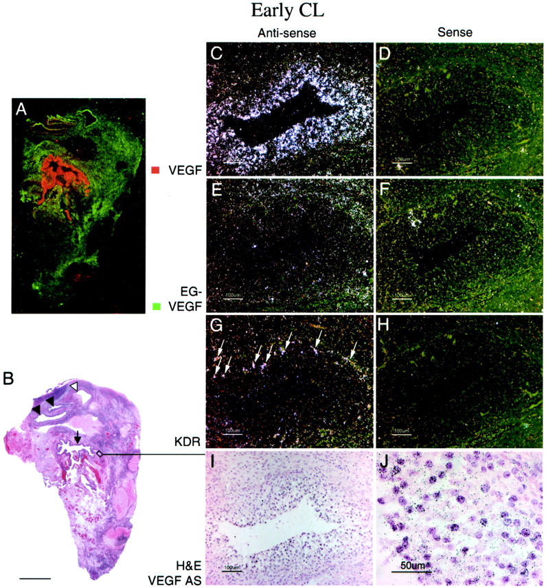
Figure 2.

EG-VEGF and VEGF expression in normal ovary early-stage CL. An early-stage (approximately day 2 to 3 after ovulation) CL, characterized by incompletely developed vascularity in the granulosa lutein layer and by inapparent theca lutein cell differentiation (I, J), shows strong VEGF expression in the granulosa lutein cells. A: False-colored autoradiographic film results show intense VEGF expression (red) in the wall of the large cystic CL (B, arrow). Microscopic results show granulosa lutein cells are intensely VEGF-positive (C, dark field; J, bright field), but only weakly positive for EG-VEGF (E); the surrounding theca is only weakly positive for both VEGF and EG-VEGF. VEGFR-2 (KDR) expression (G) is present in small vessels at the boundary between the theca interna and granulosa cell layer, and in vessels invading the outermost granulosa cell layers (I, arrows). Other atretic follicles (A, B) with (closed arrowheads) and without (open arrowhead) intact granulosa cell linings (detail not shown) show prominent EG-VEGF expression in the theca interna. Scale bars: 5 mm (B); 100 μm (C–I); 50 μm (J).
