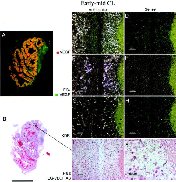
Figure 3.

EG-VEGF and VEGF expression in normal ovary early- to mid-stage CL. A CL at approximately day 5 after ovulation, characterized microscopically by well-developed vascularity and early theca lutein cell differentiation (I), shows strong VEGF (C) and EG-VEGF (E) expression in spatially overlapping CL cell populations. A: False-colored autoradiographic film results show yellow-orange overlapping of VEGF (red) and EG-VEGF (green) expression in a large cystic CL. It was not possible to determine from this result whether VEGF and EG-VEGF were co-expressed in the same cells, or in separate cells in the CL. The distribution of EG-VEGF and VEGF signal across the entire CL is consistent with expression by theca granulosa cells, but theca lutein cells may also express EG-VEGF at this stage. Vascular VEGFR-2 (KDR) expression is intense in the CL (G). Scale bars: 5 mm (B); 100 μm (C–I); 50 μm (J).
