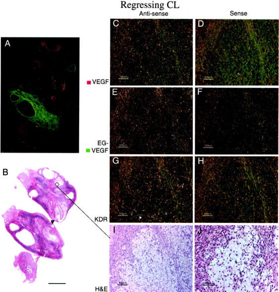
Figure 5.

EG-VEGF and VEGF expression in normal ovary late-regressing CL. A regressing CL (approximately day 14 after ovulation), characterized by large, pale, vacuolated theca granulosa and theca lutein cells (I, J), shows absence of both VEGF (C) and EG-VEGF (E) expression. A: False-colored autoradiographic film results show absence of VEGF (red) and EG-VEGF (green) signal in an area that microscopically corresponds to the regressing CL. Only weak VEGFR-2 (KDR) expression (G) is noted in scattered vessels in the granulosa cell layer. A developing tertiary (antral) follicle (A and B, arrowhead) shows strong VEGF expression (see Figure 1 ▶ for details). Scale bars: 5 mm (B); 100 μm (C–I); 50 μm (J).
