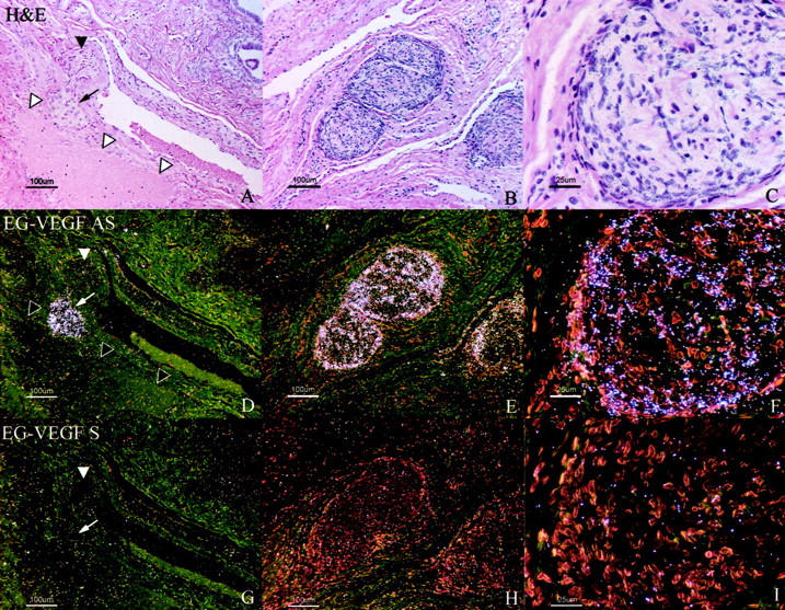
Figure 6.

EG-VEGF expression in ovarian hilus cells. EG-VEGF expression (D–F) is very strong in cells, morphologically and biochemically similar to testicular Leydig cells, found in the ovarian hilus in association with blood vessels and unmyelinated nerves. A, D, G: H&E-stained section (A) shows a nest of hilus cells (small arrow) adjacent to a large vein (open arrowheads). In this example, an adjacent unmyelinated nerve fiber (filled arrowhead) lacks associated hilus cells. B, C, E, F: Hilus cells are intimately associated with a large unmyelinated nerve fiber in this section. Epithelium of the rete ovarii (glandular structures in A, top right) expresses neither VEGF nor EG-VEGF. Scale bars: 100 μm (A, B, D, E, G, H); 25 μm (C, F, I).
