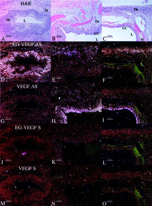
Figure 8.

Distribution of VEGF and EG-VEGF mRNA in parallel sections of cysts in individual PCOS ovaries. Parallel section were hybridized with EG-VEGF anti-sense (D–F), VEGF anti-sense (G–I), EG-VEGF sense (J–L), and VEGF sense (M–O) riboprobes. H&E images (A–C) are shown for reference. A, D, G, J, M: Detail of late-stage atretic follicle from Figure 7A ▶ , boxed area 1; EG-VEGF (D) is strongly expressed in theca cells surrounding follicle lumen in which the granulosa cell layer has degenerated. B, E, H, K, N: Detail of early-stage atretic follicle from Figure 7A ▶ , boxed area 2; VEGF (H) is strongly expressed in granulosa cells surrounding the follicle lumen; some surrounding thecal cells are weakly VEGF-positive; EG-VEGF (C) is expressed in clusters of surrounding thecal cells. C, F, I, L, O: detail from Figure 7D ▶ , boxed area, of two atretic follicles at different stages of degeneration; VEGF (I) is strongly expressed in granulosa cells surrounding lower follicle lumen; the surrounding thin layer of thecal cells are weakly VEGF-positive, and EG-VEGF-negative; EG-VEGF (F) is expressed in the thecal cells of the upper follicle in which the granulosa cell layer has degenerated. Granulosa cells (GC), theca (Th), stroma (St), lumen (L). Scale bars: 200 μm (A, D, G, J, M); 100 μm (B, C, E, F, H, I, K, L, N, O).
