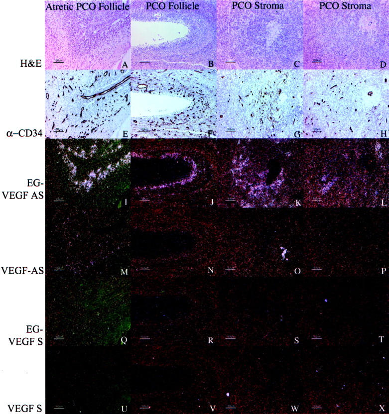
Figure 9.

Correlation between expression of VEGF or EG-VEGF and vascularity, as assessed by expression of CD34, in representative PCOS specimens. Parallel sections were immunostained with anti-CD34 (QBEnd/10, E–H) or hybridized with EG-VEGF anti-sense (I–L), VEGF anti-sense (M–P), EG-VEGF sense (Q–T), and VEGF sense (U–X) riboprobes. H&E images (A–D) are shown for reference. In PCOS ovaries, EG-VEGF expression is high in the theca surrounding atretic follicle lumens (A, B, I, J) and diffusely in ovarian stroma (C, D, K, L), whereas VEGF expression in these areas (Q–T) is weak or absent. Vascularity in corresponding areas is illustrated by CD34 immunostaining (E–H). Similar, although weaker immunostaining was observed with anti-CD31 monoclonal antibody JC/70A (not shown). Scale bars, 100 μm.
