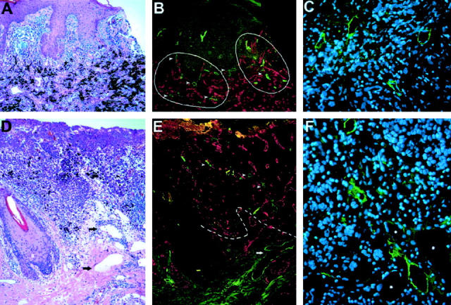
Figure 1.

Detection of intratumoral and peritumoral lymphatic vessels in cutaneous malignant melanomas. A: Histology of a thick melanoma (4.5 mm) shows compact masses of frequently pigmented tumor cells. B: Immunofluorescent stain of a serial section of the same tumor for the lymphatic marker LYVE-1 (green) and the panvascular marker CD31 (red) reveals prominent hotspots of high lymphatic vessel density (hotspots are circled by solid line). In contrast, blood vessels are homogeneously distributed throughout the tumor. C: Thin-walled, LYVE-1-positive intratumoral lymphatic vessels with open lumina. D: Histology of a thin melanoma (≤1.5 mm) with dilated peritumoral lymphatics (arrows). E: Immunofluorescent stain of a serial section for LYVE-1 (green) and CD31 (red) reveals lymphatic vessels within (arrowheads) and surrounding (arrows) the tumor border (dotted line). F: LYVE-1-positive intratumoral lymphatic vessels with open lumina. The adjacent blood vessels (asterisks) are LYVE-1-negative. Cell nuclei are counterstained (blue) with Hoechst (C and F). Original magnifications: ×100 (A, B, D, and E); ×400 (C and F).
