Figure 2.

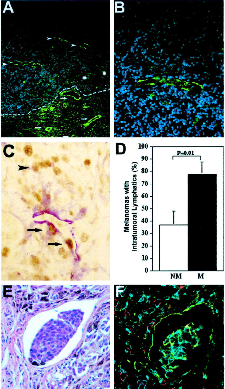
Higher frequency of intratumoral lymphangiogenesis in metastatic melanomas. A: Immunofluorescent stain for LYVE-1 (green) depicts thick-walled peritumoral (arrows) and thin-walled intratumoral lymphatics (arrowheads) in a thin melanoma (1.05 mm). The tumor border is indicated by a dotted line. Blood vessels (asterisks) are negative for LYVE-1. B: Higher magnification of intratumoral lymphatics reveals thin-walled, basket-like morphology. C: Double immunostain for LYVE-1 (red) and PCNA (brown) reveals an intratumoral lymphatic vessel with proliferating lymphatic endothelial cells (arrows) and adjacent melanoma cells (arrowhead). D: Significantly increased frequency of detectable intratumoral lymphatic vessels in metastatic melanomas (M; n = 18), as compared with nonmetastatic (NM; n = 19) tumors (mean ± SEM, chi-square test, P = 0.01). E: Detection of melanin-containing tumor cells within an intratumoral lymphatic vessel. H&E stain. F: Immunofluorescent stain of a serial section for LYVE-1 (green) and CD31 (red) confirms that tumor cells are located within a lymphatic vessel. Cell nuclei are counterstained blue with Hoechst (B and F). Original magnifications: ×200 (A); ×400 (B, C, E, F).
