
Figure 3.
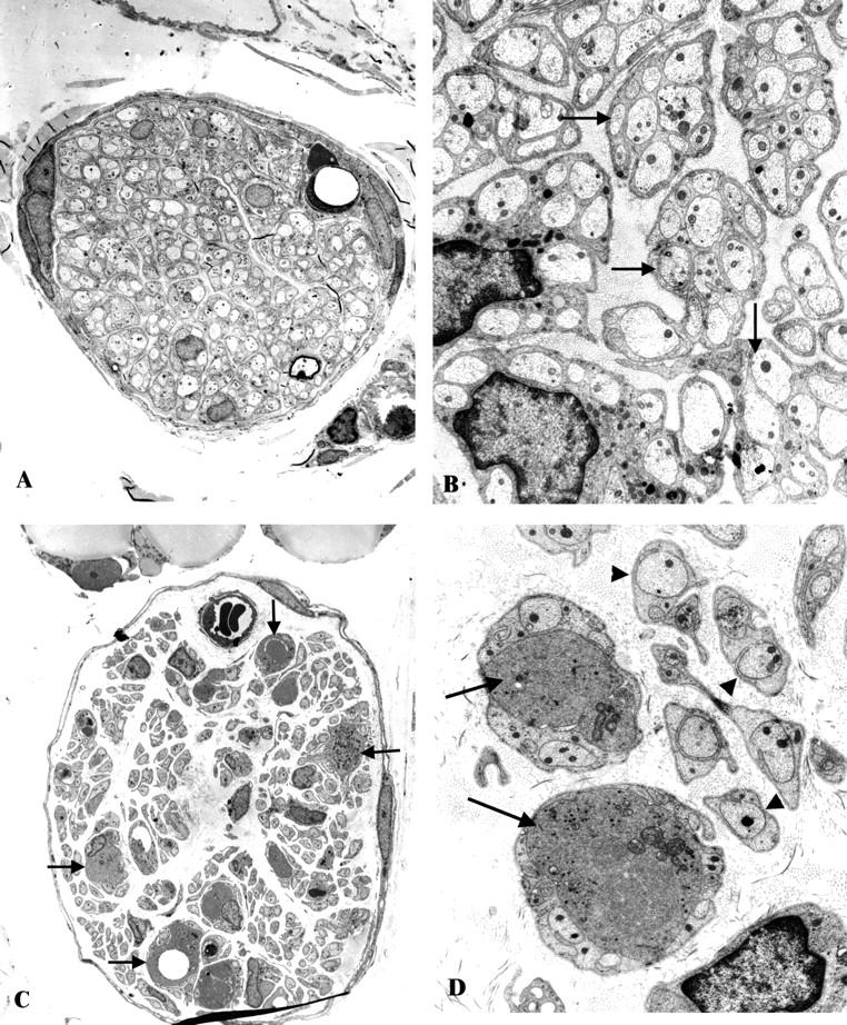
Neuroaxonal dystrophy in the ileal mesenteric nerves of ZDF- and STZ-diabetic rats. A and B: A typical mesenteric nerve fascicle in the ZDF rat (A) contains numerous normal appearing unmyelinated axons (arrows, B) without significant numbers of dystrophic axons. (Original magnification: A, ×1200; B, ×7500). C and D: A single mesenteric STZ-diabetic rat nerve fascicle (C) shows numerous unmyelinated axons (arrowheads, D) some of which demonstrate the ultrastructural appearance of neuroaxonal dystrophy (arrows, C and D). (Original magnification: C, ×1200; D, ×7500)