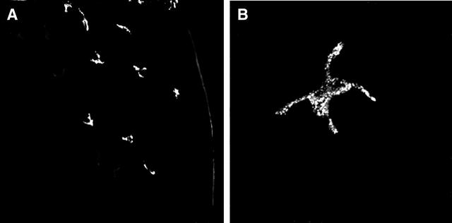
Figure 1.
VEGFR-3 expression on cells in the periphery of the corneal stroma. Confocal micrographs demonstrate VEGFR-3+ dendritic shaped cellular structures in the periphery (right) of the cornea, while the central areas (left) do not demonstrate such cells (A). Higher magnification, confirming the dendritic morphology of VEGFR-3+ cells in the cornea (B). Magnification, (A) ×200, (B) ×1000.

