
Figure 2.
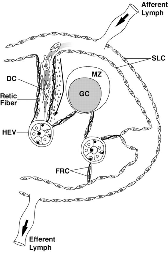
A model of the lymph node reticular network extending from SLC to the HEV. At the upper left, an expanded view of the proposed structure is shown with a dendritic cell (DC) migrating from the subcapsular sinus through a corridor created by reticular fibers lined by FRC. Solute, represented as small dots, is shown spreading down a conduit between the FRC and the reticular fiber. The reticular network does not extend into the GC (Adapted from the model of Anderson and Shaw).