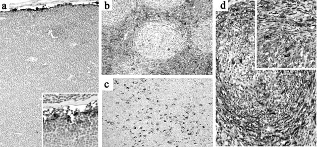
Figure 6.
Tissue TG staining in lymphoma. a: Small lymphocytic lymphoma show a virtual absence of staining in HEV and the FRC network with strong TG immunoreactivity limited to SLC (inset). b: Follicular lymphoma with strongly TG+ FRC network, blood vessels and dendritic cells surrounding neoplastic follicles. c: Mantle cell lymphoma shows numerous TG+ histiocytoid cells with the FRC network negative for TG. d: Nodular sclerosis Hodgkin’s disease show intensely TG+ myofibroblasts in perinodular areas extending into adjacent uninvolved parenchyma (inset).

