
Figure 1.
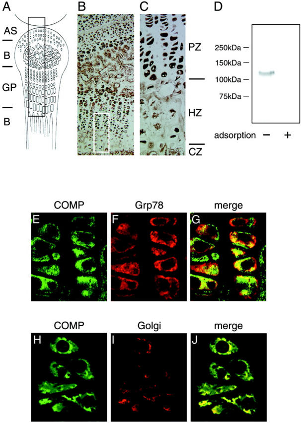
Characterization of a newly-prepared anti-COMP antibody. Murine growth plates of the knee including chondrocytes at 2 weeks after birth were fixed and embedded in paraffin. Sections were stained with a newly prepared anti-COMP antibody and detected by diaminobenzidine and 0.03% hydrogen peroxide. A: Schema of sagittal section of murine knee joint including a growth plate (GP). AS and B represent articular surface and bone, respectively. B: Stained section of murine knee joint with anti-COMP antibody that represents the black boxed area in A (Magnification, ×10). C: The white boxed area in B at higher magnification (Magnification, ×40). Note that the distribution of immunoreactivity with this antibody was similar to that with a COMP-specific antibody described in a previous study. 40 PZ, HZ, and CZ represent the proliferating zone, hypertrophic zone, and calcification zone, respectively. D: Western blot showing that anti-COMP antibody specifically recognized native COMP protein synthesized in chondrocytes. The symbols (+) and (-) indicate with or without recombinant COMP fragment used for the adsorption experiment as described in Materials and Methods. E–J: Confocal view to examine the subcellular distribution of COMP in chondrocytes of cartilage. The cartilage tissue section was stained with anti-COMP antibody (E, H) and anti-ER antibody (F) or anti-Golgi antibody (I) followed by relevant FITC- and rhodamine-labeled secondary antibodies. G and J are merged images of E and F, and H and I, respectively. Experimental details are provided in Materials and Methods.