
Figure 5.
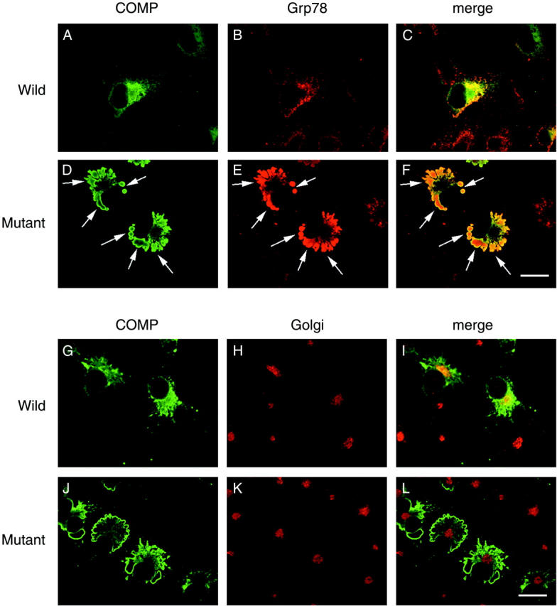
Confocal analysis of subcellular localization of COMP. COS7 cells were transfected with wild-type (A, B, C, G, H, and I) or mutated COMP (D, E, F, J, K, and L) and were cultured for 48 hours at 37°C. The cells were fixed and incubated with the primary antibodies described below followed by FITC- and rhodamine-labeled secondary antibodies. Primary antibodies were the anti-COMP antibody (A, D, G, and J), an anti-Grp78 (an ER maker) antibody (B and E) and an anti-Golgi 58K protein (a Golgi apparatus maker) antibody (H and K). C and F are merged images of COMP and Grp78 staining, while J and L are those of COMP and Golgi 58K protein staining. Virtually all COMP staining in cells with mutated COMP overlapped Grp78 staining (arrows). No COMP staining was detected in untransfected cells. Wild-type COMP was partially colocalized with the Golgi apparatus, whereas mutated COMP was not associated with the Golgi apparatus. Bar, 20 μm.