
Figure 2.
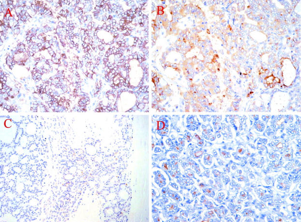
A and B: Consecutive serial sections of a follicular variant of PTC stained with mAbs HBME-1 (A) and 373E1 (B), respectively. Note how the former shows a cell membrane-associated staining contrasting the diffuse cytoplasmic and apical ones observed with mAb 373E1. C and D: Direct comparison of the staining patterns observed with 373E1 in a follicular carcinoma (C), showing tumor capsule penetration, and in a follicular variant of PTC (D).