
Figure 4.
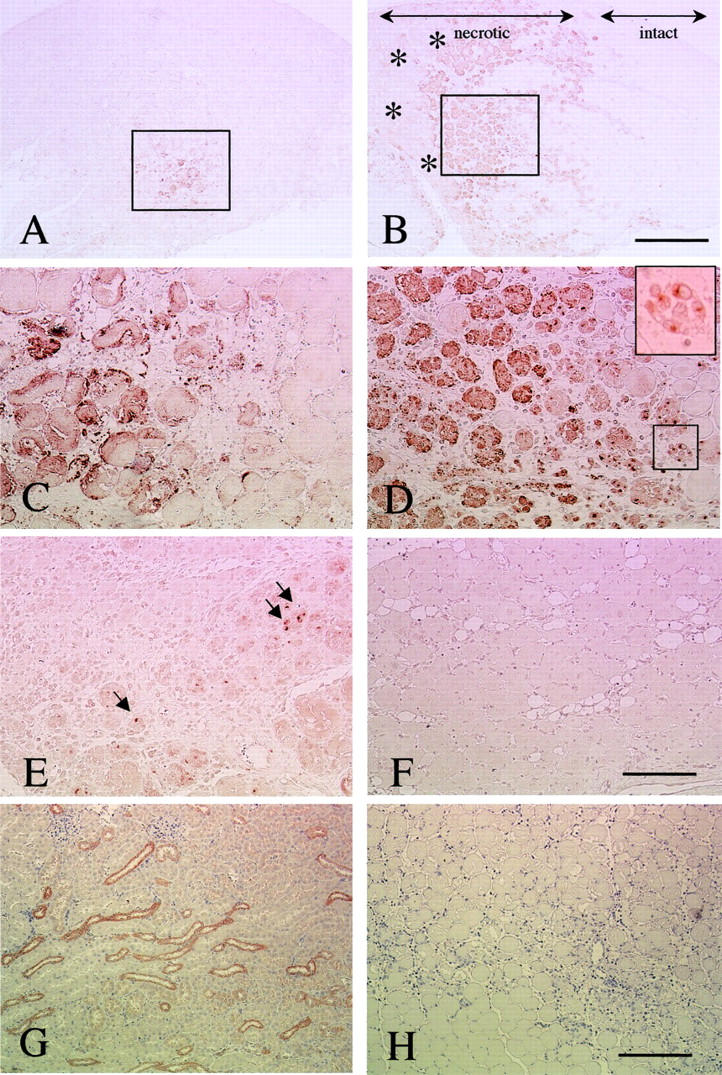
Immunohistochemical analysis of OPN in cardiotoxin-treated muscle. Transverse sections at 24 hours (A, C), 48 hours (B, D), 96 hours (E), and 7 days (F) after cardiotoxin injection were stained with anti-OPN antibody. C and D are high-power magnifications of the areas indicated by rectangles in A and B, respectively. The immunoreactivity is observed in mononucleated cells (inset in D) and necrotic fibers. The intact area, which escaped cardiotoxin injury, is indicated in B. Note that necrotic areas where mononucleated cells have not infiltrated yet were negative for OPN staining (asterisks in B). At 96 hours after treatment, OPN expression was considerably decreased when compared to that at 48 hours, but some OPN-positive mononucleated cells were observed (arrows in E). F: OPN staining was not detected at 7 days after the treatment. G: Positive control section from kidney of ICR mice. OPN was expressed in the long loop of Henle and in the distal tubule of the kidney. H: The negative control section of muscle at 2 days after cardiotoxin treatment showed no apparent staining. All sections were stained by the immunoperoxidase (ABC) method with methyl green (A-F) or hematoxylin (G and H). Scale bars: 400 μm (A, B), 200 μm (C, D, E, and B), 100 μm (G and H).