
Figure 2.
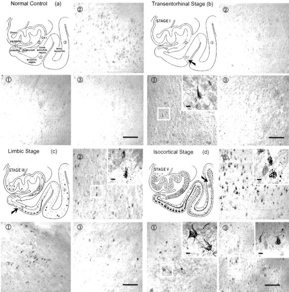
Immunohistochemical staining with antibody to activated p70 S6 kinase (T389) in brains with different degrees of neurofibrillary degeneration according to Braak’s staging criteria. The normal control that does not have any neurofibrillary involvement did not reveal any immunostaining (a). The early transentorhinal stage (I-II) that is characterized by mild neurofibrillary pathology in the transentorhinal region showed a few immunopositive neurons (b). The moderate limbic stage (III-IV) which is marked by a moderate involvement of neurofibrillary pathology in the entorhinal region, and the involvement of a few or many CA1 cells in the hippocampal region and temporal lobes showed immunostaining in several tangle-bearing neurons (c). The late isocortical stage (V-VI) which is characterized by the involvement of severe neurofibrillary pathology in the entorhinal cortex, the hippocampus, and isocortex showed the most robust immunostaining of neurons (d). Following the sequence of neurofibrillary degeneration from normal control to isocortical stage (V/VI), antibody to p70 S6 kinase phosphorylated at T389 showed progressively increased numbers of tangle-like inclusions and granular structures. Area 1 shows transentorhinal region in a and b or entorhinal region in c and d. Areas 2 and 3 show hippocampal CA1 and temporal isocortex, respectively in a, b, c and d. Bars in 3, 100 μm; bars in insets, 10 μm.