
Figure 5.
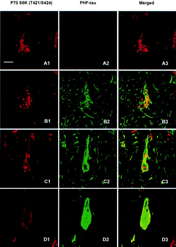
Immunoreactivity of active P70 S6 kinase (T421/S424) in neurons with different degrees of AT8-labeled PHF-tau involvement. In the normal-looking neurons, only active p70 S6 kinase was positive in granular form (A1, A2, and A3). In the pre-tangle neurons, some of the granular stainings of active p70 S6 kinase was partially overlapped with dotted AT8 labeled PHF-tau (B1, B2, and B3). In classic tangle bearing neurons, the granular staining of active p70 S6 kinase congregated, and some particle staining distributed along with the AT8-positive filamentous structures (C1, C2, and C3). At the advanced stage of tangle neurons, no dot-like active p70 S6 kinase staining could be seen, except for the faint filamentous structures partially overlapping with AT8 positive tangles (D1, D2, and D3). Bars, 10 μm.