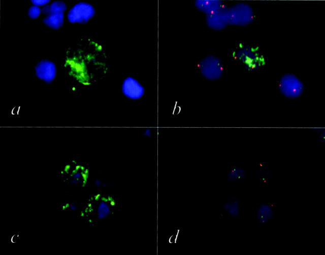
Figure 2.
a and c: Bone marrow cells from a neuroblastoma patient stained with FITC labeled GD2 antibody. b and d: After automatic relocation of the GD2-positive cells these cells were subsequently analyzed by FISH using a MYCN specific probe (FITC) and a chromosome 2-specific probe (TRITC). Only the GD2-positive cell shown in a and b displays the tumor typical MYCN amplification (b). The three GD2-positive cells shown in c show a normal MYCN copy number (d). Applying this genetic verification procedure (AIPF), tumor cells can be unambiguously discriminated from normal cells.

