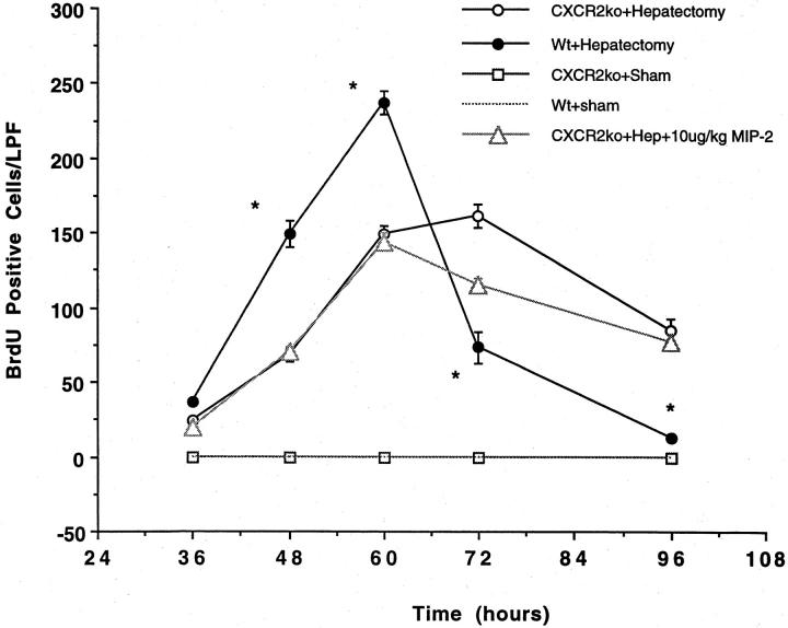
Figure 4.
BrdU staining in CXCR2 knockout mice or wild-type controls undergoing partial hepatectomy. Partial hepatectomy was performed in CXCR2 knockout or wild-type control mice. An additional group of CXCR2 knockout mice were treated with 10 μg/kg of MIP-2 and underwent hepatectomy. BrdU staining was performed on liver tissue obtained at 36, 48, 60, 72, and 96 hours after resection and is expressed as the number of BrdU-positive cells per low-power field (LPF). There was a significant decrease in the number of BrdU-positive cells in the CXCR2 knockout mice at 48, 60, 72, and 96 hours after resection, as compared to wild-type mice; this difference was not corrected by administration of exogenous MIP-2, suggesting that MIP-2 functions in this system via the CXCR2 receptor. *, P < 0.05 versus CXCR2ko + Hep and CXCR2ko + 10 μg/kg MIP-2.

