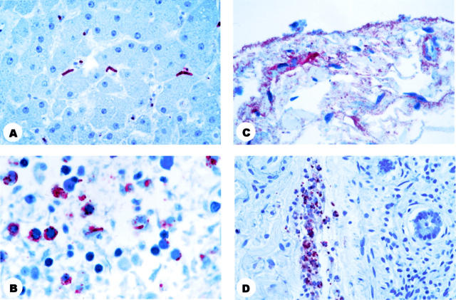
Figure 4.
Photomicrographs showing immunohistochemistry of abdominal organs from patients who died. A: Liver showing bacilli in the sinusoids and bacillary fragments in Kupffer’s cells. B: Spleen showing bacillary fragments and granular antigen-staining. C: Intestinal serosa showing granular antigen-staining. D: Intestinal submocosal blood vessel showing granular antigen-staining. Immunohistochemical assay using the B. anthracis capsule antibody (B, C, and D) or the B. anthracis cell wall antibody (A) naphthol/fast red substrate with hematoxylin counterstain. Original magnifications: A and B, ×100; C, ×63; D, ×40.

