
Figure 1.
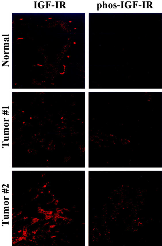
Immunohistochemical staining for IGF-IR and activated IGF-IR in human pancreatic cancers and nonmalignant pancreatic tissues. Paraffin-embedded specimens from human pancreatic cancers and nonmalignant tissues were immunofluorescently stained for IGF-IR and phosphorylated IGF-IR. IGF-IR was expressed in normal ductal epithelium as well as tumor epithelium. However, only tumor epithelium stained positive for phosphorylated IGF-IR.