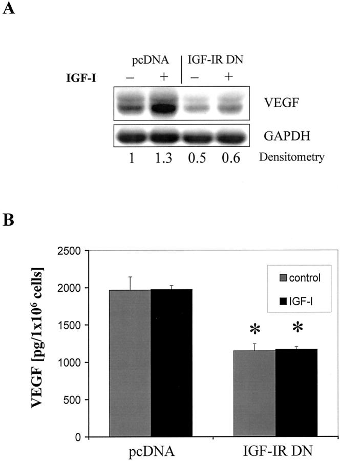
Figure 3.
Effect of IGF-IR inhibition on constitutive and inducible VEGF expression. A: Northern blot analysis of VEGF expression in pcDNA- and IGF-IR DN-transfected L3.6pl cells after a 24-hour incubation in serum-reduced conditions (1% FBS-MEM) with or without rhIGF-I (100 ng/ml). Relative changes in VEGF mRNA expression with respect to controls (unstimulated pcDNA cells) are shown beneath each lane. B: ELISA of VEGF protein concentration in conditioned medium of transfected L3.6pl cells. Medium was collected after a 48-hour incubation period in 10% FBS-MEM in the presence or absence of rhIGF-I (100 ng/ml). *, P < 0.01 versus treated and untreated pcDNA cells, Student’s t-test. Data are presented as means ± SEM.

