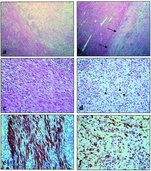
Figure 2.

Overview of the histopathological and immunohistochemical findings. In many areas, the transition between the smooth-muscle component (left) and the IPT component (right) is easily seen (H&E stain, ×25) (a). The tumor is relatively well delineated from the surrounding parenchyma (arrows) (H&E stain, ×25) (b). Higher magnification of the smooth-muscle component shows bundles of eosinophilic spindle cells with cigar-shaped nuclei (H&E stain, ×160) (c). Higher power of the IPT component discloses vessels, lymphocytes, plasma cells (arrowheads), and spindle cells in a myxoid background (H&E stain, ×160) (d). The smooth-muscle component is strongly desmin-positive (indirect immunoperoxidase stain, ×400) (e). The spindle cells of the IPT component only express α-SMA (indirect immunoperoxidase stain, ×400) (f).
