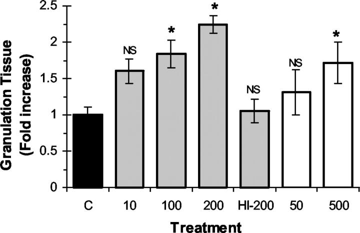
Figure 2.
Effect of recombinant EPO on granulation tissue formation during wound healing. F-ZCs were harvested at day 6 after implantation and granulation tissue formation was measured as described in Materials and Methods. The granulation tissue thickness in each treatment group was expressed as fold change from control (y axis). mEPO (gray bars) was administered into F-ZCs at the indicated concentrations ranging from 10 to 200 U/ml and hEPO (white bars) was administered at concentrations of 50 and 500 U/ml. The negative control chambers (C) contained vehicle (black bar) or 200 U/ml of heat-inactivated mEPO (HI-200). The mean values (±SE) are shown. *, P < 0.05 (analysis of variance). NS, not significant. Table 1 ▶ illustrates the measurement data, number of measurements in each group, and the P values.

