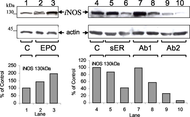
Figure 5.
Immunoblot for iNOS expression in granulation tissue during wound healing. Granulation tissues from F-ZCs were harvested at day 6 after administration of EPO (left, lanes 1 to 3) or day 9 after soluble EPOR (sER), mAb 2871 (Ab1), or mAb 287 (Ab2) administration (right, lanes 4 to 10). Western blotting was performed as described in Materials and Methods. Lane 1, control day 6 (C); lane 2, EPO 50 U/ml; lane 3, EPO 500 U/ml; lane 4, control day 9 (C); lane 5, sER 1 μg/ml; lane 6, sER 10 μg/ml; lane 7, Ab1 10 μg/ml; lane 8, Ab1 100 μg/ml; lane 9, Ab2 5 μg/ml; and lane 10, Ab2 50 μg/ml. Comparable loading and integrity of proteins in each lane were confirmed by hybridization of an immunoblot to anti-actin antibody (bottom blots). Arrows indicate iNOS and actin immunoreactivity. Densitometric analysis for the 130-kd iNOS bands for this representative experiment are illustrated in the graphs below the blots.

