
Figure 4.
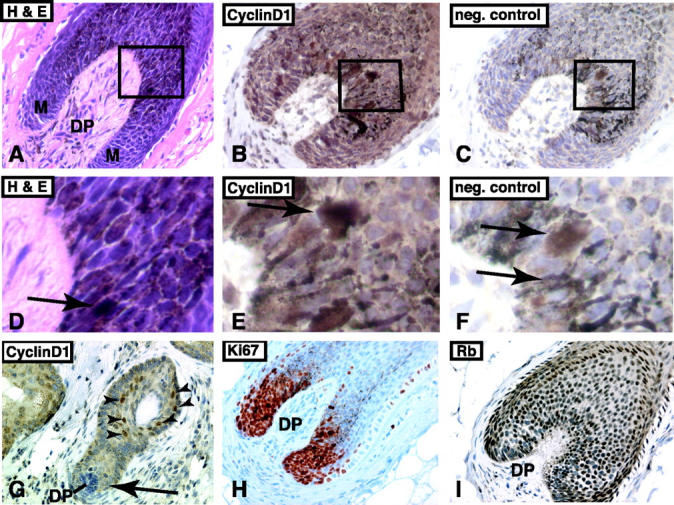
Immunohistochemical detection of cyclin D1, Ki67, and RB in the anagen hair bulb. A: H&E-stained tissue section of anagen hair bulb (DP, dermal papilla; M, matrix; ×200). Higher magnification view of boxed area is shown in D. B: Immunohistochemical staining for cyclin D1 in anagen bulb (×400). The anagen bulb is rich in pigment-laden melanocytes. No true nuclear staining is present. The boxed area is shown in E. C: Negative control of consecutive section of anagen bulb shown in B. The pigment-laden melanocytes are easily identified (×400). The boxed area is shown in F. Arrows in D, E, and F point to pigment-laden cells. G: At anagen onset, the suprabasal cells of the telogen bulge are positive for cyclin D1 (arrowheads). However, the newly formed anagen bulb is negative for cyclin D1 (arrow, ×200). H: Ki67 staining shows that cells around the dermal papilla are strongly positive and proliferating (×200). E: Immunostaining for Rb shows that the matrix cells retain expression of Rb (×200). Immunohistochemical sections are counterstained with hematoxylin.