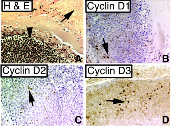
Figure 7.

Pilomatricomas express cyclins D1, D2, and D3. A: H&E stain of pilomatricoma. Arrow indicates shadow cells; arrowhead indicates basoloid cells (×200), Tumor cells are focally positive for cyclin D1 (B), cyclin D2 (C), and cyclin D3 (D) (arrows, ×200).
