Figure 3.

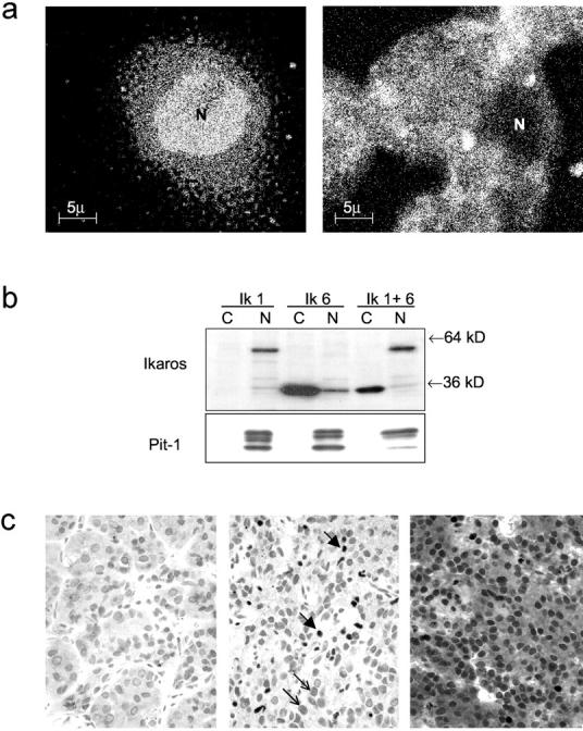
Localization of Ik1 and Ik6 in pituitary cells. a: Immunofluorescence detection of Ik isoforms. GH4 pituitary cells were transiently transfected with Ik1 or Ik6. Note the predominantly nuclear (N) staining in Ik1-transfected cells (left) in contrast to the cytoplasmic fluorescence detected in Ik6-transfected cells (right). b: Western blotting of fractionated protein lysates from Ik-1-transfected GH4 cells detects a 57-kd protein predominantly in nuclear fractions (N); Ik-6-transfected cells exhibit a 36-kd protein predominantly in cytoplasmic fractions (C) with some component in nuclear fractions (N). Cells transfected with both Ik1 and Ik6 (Ik 1 + 6) show nuclear Ik1 and predominantly cytoplasmic Ik6 reactivity. Pit-1 reactivity was used as a positive control to verify appropriate fractionation of protein. c: Immunocytochemical localization of Ik in the human pituitary. The nontumorous adenohypophysis (left) is negative. A pituitary adenoma that expressed Ik1 (middle) exhibits focal strong nuclear staining (thick arrows). An adenoma that expressed Ik1 and Ik6 (right) exhibits strong nuclear and cytoplasmic staining.
