
Figure 1.
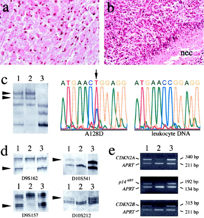
a and b: Histological features of the primary and recurrent glioma of patient 2 on hematoxylin-eosin stained sections. a: The primary tumor (A128D) was a gemistocytic astrocytoma (WHO grade II). b: The recurrent tumor (GB119D) displayed increased cellularity, high mitotic activity, microvascular proliferation, and pseudopalisading necroses (nec). This tumor was classified as glioblastoma (WHO grade IV). c to e: Examples of molecular genetic results obtained for these tumors (lane 1, A128D; lane 2, GB119D; lane 3, leukocyte DNA from patient 2). c: SSCP analysis of TP53 showed identical aberrant band patterns in A128D and GB119D. DNA sequencing revealed the same somatic TP53 mutation (g.14070C>T; R248W) in both tumors. Shown are the mutant sequence in A128D and the wild-type sequence in the corresponding leukocyte DNA. d: Microsatellite analysis of polymorphic loci from 9p (D9S162 and D9S157) revealed LOH only in GB119D but not in A128D. LOH on 10q was detectable in both tumors, as indicated for the loci D10S541 and D10S212. However, the fraction of tumor cells with LOH on 10q appeared to be higher in GB119D than in A128D (arrowheads point to the alleles lost in the tumor DNA). e: Duplex PCR analysis demonstrated homozygous deletion of CDKN2A, p14ARF and CDKN2B in GB119D but not in A128D. The sizes of the respective PCR fragments amplified from each of these genes and the reference gene APRT are indicated on the right side.